

 en
en
 English
 繁体中文
繁体中文 Japanese
 Korean
 Russian
 German
News
Deepen Optoelectronic Resources, Lead Technological Breakthroughs

Deepen Optoelectronic Resources, Lead Technological Breakthroughs
3D endoscope technology has become one of the indispensable and important tools in modern medicine. It enables doctors to observe and manipulate organs and tissues in the patient's body in real-time during surgery by utilizing advanced optical and imaging technologies.
1. Working principle of 3D endoscope
1. Optical principle:
The 3D endoscope uses optical imaging technology. It consists of a small optical lens, fiber optic bundle, camera, and display screen. The optical lens transmits light to the camera through a fiber optic bundle, which converts the light into electrical signals and transmits them to the display screen for real-time observation.
2. Principle of Stereoscopic Imaging:
The 3D endoscope utilizes the principle of stereoscopic imaging to observe three-dimensional images. It has two cameras installed on the optical lens, which are at a certain distance. By combining the images captured by two cameras, a stereoscopic image can be obtained, allowing doctors to observe the location and morphology of lesions more clearly.
2. The application of 3D endoscope
1. Laparoscopic surgery:
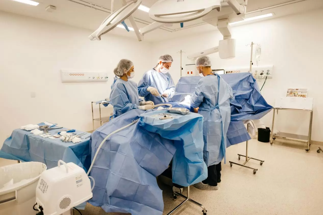
3D endoscopy has been widely used in laparoscopic surgery. Traditional laparoscopic surgery uses a 2D endoscope, and doctors can only observe flat images, making it difficult to accurately determine the depth and distance of organs. 3D endoscopes can provide more realistic and clear stereoscopic images, enabling doctors to perform surgical operations more accurately and reduce surgical risks.
2. Thoracoscopic surgery:
3D endoscopy also has important applications in thoracoscopic surgery. Thoracoscopic surgery requires operation in a very narrow space, and traditional 2D endoscopes cannot provide sufficient spatial perception ability. 3D endoscopes can provide more three-dimensional images, allowing doctors to more accurately locate and manipulate the affected area, improving the success rate and safety of surgery.
3. Gastroscopy examination:
Gastroscopy is one of the common endoscopic examinations. Traditional gastroscopy uses a 2D endoscope, and doctors can only observe flat images, which has certain limitations in the diagnosis of gastric lesions. 3D endoscopes can provide more realistic and clear stereoscopic images, allowing doctors to more accurately determine the location and degree of lesions and improve diagnostic accuracy.
The emergence of 3D endoscope technology has brought revolutionary changes to the medical field. It enables doctors to observe and manipulate organs and tissues in the patient's body in real-time during surgery by utilizing advanced optical and imaging technologies. 3D endoscopy has been widely used in fields such as laparoscopic surgery, thoracoscopy, and gastroscopy, providing doctors with more accurate and safe surgical procedures and diagnostic judgments. With the continuous development of technology, it is believed that the application of 3D endoscope technology in the medical field will become increasingly widespread, bringing better treatment effects to patients.
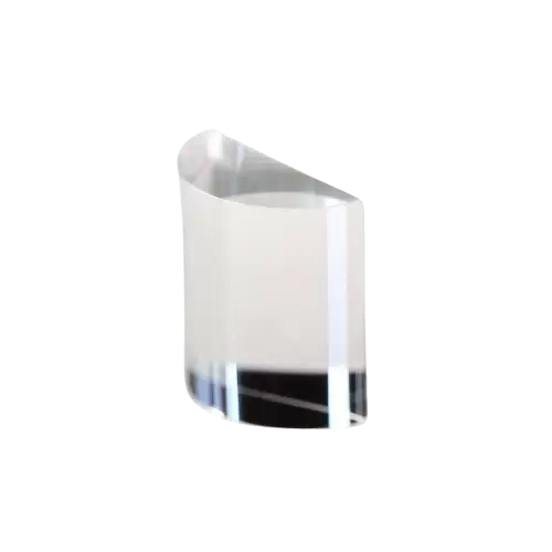
In modern optical engineering, plano convex lenses are no longer treated as simple focusing elements. Instead, they are engineered components that directly determine energy distribution accuracy, wavefront integrity, and system-level optical efficiency in applications ranging from high-power laser processing to precision metrology and scientific imaging systems.
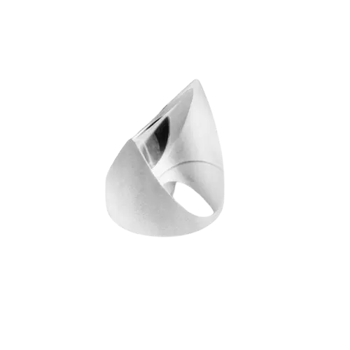
When engineers evaluate beam-shaping components for machine vision, laser measurement, industrial alignment, or optical inspection systems, the discussion quickly moves beyond simple beam expansion.
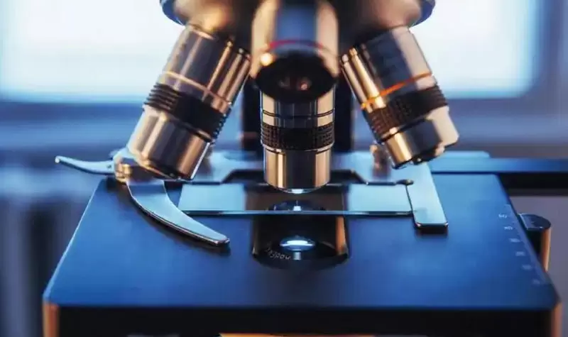
A microscope is a precision measuring instrument that allows students to observe extremely small organisms and study their microbial morphology. The main body of the microscope is composed of optical and mechanical parts, among which the optical part is the most important. The optical part of a microscope mainly consists of an objective lens and an eyepiece. By combining the two, a good observation image is formed.